
Medical Image Segmentation and Screening AI (Radiology)
Models that outline organs and lesions or flag disease on X-ray, CT, MRI and other scans.
Tier Grades
Tier A: Frontier AI labs
feasible
Big tech and hospital labs release medical encoders and segmenters.
Tier B: Indonesian AI labs
feasible
An Indonesian team built TB screening on local data (UGM), so local training is feasible.
Tier C: Institution units such as ITB
feasible
A 6-author team built MedSAM from public data; a university unit can do similar work.
Tier D: Organisations such as SEMAI (PoT, PoC, PoV)
stretch
Clinical pilots need hospital partners and regulatory review, so delivery is a stretch.
Where this comes from
Tiers: a) frontier AI labs, b) Indonesian AI labs, c) institution units such as ITB, d) organisations such as SEMAI delivering proof of technology, concept and value. Each rationale cites the anchors below.
Anchors
- ComputeTraining compute is not in the sources read.
- DataMedSAM uses 1,570,263 image-mask pairs across 10 imaging modalities and over 30 cancer types.
- Team size (proxy)Proxy only (author count of an exemplar paper, not a headcount): 6 authors on the MedSAM paper (arXiv 2304.12306).
- Time horizonYear.
- Shelf-life signalsPromptable medical segmenters replace per-task models, National TB and cancer screening programmes adopt AI readers
- Pipeline roleTier A and hospital labs release models; Tier C and Tier B validate them on Indonesian scans; Tier D supports screening pilots with clinical partners.
Where this comes from
Team size is a proxy (author counts of exemplar papers), not a headcount.
Who and What
Exemplars
Observed actors
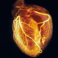 Bo Wang Lab, Toronto
 DKFZ (German Cancer Research Center)
 Helmholtz Imaging
 Qure.ai
 Universitas Gadjah Mada (UGM)
 University of Melbourne
Linked models
MedSAMvisionexemplarnnU-Net v2visionexemplarqXRvisionexemplarTBScreen.AIvisionexemplarMedSAM2visionproduced by MedVAEvisionproduced by
 CXR Foundationvisionproduced byDerm Foundationvisionproduced by
 MedImageParse (BiomedParse)visionproduced by
 MAISI (Medical AI for Synthetic Imaging)image generationproduced byRAD-DINOvisionproduced by
 UNet++visionproduced byCheXNetvisionproduced by
 U-Netvisionproduced byU-Net (2D)visionproduced by
 CAD4TBvisionproduced byMedSigLIPvisionproduced by
 Lunit INSIGHT CXRvisionproduced byLunit INSIGHT MMGvisionproduced by
 Transparavisionproduced by
 SELENA+visionproduced by
 United Imaging pulmonary nodule softwarevisionproduced by3D U-Net (TensorFlow volumetric models)visionproduced bySAM ViT-Bvisionused by
Links
R&D TopicMedical Image Segmentation and Screening AI (Radiology)
ModelsMedSAMBo Wang Lab, Toronto, visionnnU-Net v2DKFZ (German Cancer Research Center), visionqXRQure.ai, visionTBScreen.AIUniversitas Gadjah Mada (UGM), vision+25 more
MedSAMBo Wang Lab, Toronto, visionnnU-Net v2DKFZ (German Cancer Research Center), visionqXRQure.ai, visionTBScreen.AIUniversitas Gadjah Mada (UGM), vision+25 more
Models 29
U-NetUniversity of Freiburg, visionproduced byMedSAMBo Wang Lab, Toronto, visionexemplarMedSAM2Bo Wang Lab, Toronto, visionproduced byMedSigLIPGoogle, visionproduced byTBScreen.AIUniversitas Gadjah Mada (UGM), University of Melbourne, visionexemplarUNet++Arizona State University, visionproduced by3D U-Net (TensorFlow volumetric models)Google, visionproduced bynnU-Net v2DKFZ (German Cancer Research Center), Helmholtz Imaging, visionexemplarqXRQure.ai, visionexemplarSELENA+Synapxe, visionproduced byTransparaScreenPoint Medical, visionproduced byU-Net (2D)University of Freiburg, visionproduced by+17
United Imaging pulmonary nodule softwareShanghai United Imaging, visionproduced byVISTA-2D and VISTA-3DNVIDIA, visionproduced byCAD4TBDelft Imaging, visionproduced byDerm FoundationGoogle, visionproduced byLunit INSIGHT CXRLunit, visionproduced byLunit INSIGHT MMGLunit, visionproduced byMAISI (Medical AI for Synthetic Imaging)NVIDIA, image generationproduced byMedImageParse (BiomedParse)Microsoft AI, visionproduced byMONAI model bundlesNVIDIA, visionproduced byPolyp Detection RT-DETR (Holoscan)NVIDIA, visionproduced byRAD-DINOMicrosoft AI, visionproduced bySAM ViT-Bvisionused bySigLIP-400Mvisionused byCheXNetStanford University, visionproduced byCPPE-5 medical PPE detectorsvisionproduced byCXR FoundationGoogle, visionproduced byMedVAEStanford University, visionproduced byPapers
- U-Net: Convolutional Networks for Biomedical Image SegmentationOlaf Ronneberger et al., MICCAI 2015 (per arXiv comment), 2015 founding
- Segment Anything in Medical ImagesJun Ma et al., Nature Communications 15, 654 (2024), 2023 state of the art
- TotalSegmentator: robust segmentation of 104 anatomical structures in CT imagesJakob Wasserthal et al., Radiol Artif Intell 2023;5(5):e230024, 2022 state of the art
- TransUNet: Transformers Make Strong Encoders for Medical Image SegmentationJieneng Chen et al., arXiv preprint, 2021 state of the art
- nnU-Net: Self-adapting Framework for U-Net-Based Medical Image SegmentationFabian Isensee et al., arXiv preprint, 2018 state of the art
Where this comes from
The papers to read first: where the direction began, its current best, and a survey when one exists.
Sources
- arxiv.org/abs/2304.12306 https://arxiv.org/abs/2304.12306
To cite this page: Project AURION, AI Use-Cases Research & Innovation, "Medical Image Segmentation and Screening AI (Radiology)", https://aurion.semai.id/topics/medical-image-segmentation-and-screening/, records of October 2026.